
Prof. Dr. Arnd Dörfler
Neuroradiology Department
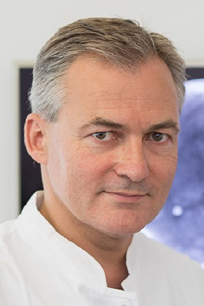
The scientific focus of the Department of Neuroradiology is on multimodal imaging, especially in stroke, brain tumors, focal epilepsies, MS and dementia, and on the minimally invasive treatment of cerebral aneurysms, vascular malformations, stenosis and acute stroke using state-of-the-art imaging technology including innovative flat-panel imaging and clinically approved ultrahigh-field 7 Tesla MRI.
Research Projects
- Department of Neuroradiology, University Hospital Erlangen, FAU
- Professorship for Neuroradiology (Chair; W3)
- Professorship for multimodal clinical imaging (W2; Prof. Zaiss)
- Multimodal imaging of cerebrovascular diseases, epilepsy, neurooncology and neurodegenerative disease
- 7 Tesla ultrahigh-field neuroimaging
- Functional and metabolic MR imaging
- Minimally invasive neurovascular and spinal interventions and interventional imaging
- AI applications in neuroimaging
J111: Voxelomic Atlas: Single-Voxel Spatio-Spectral Homology Matching
(FAU Funds)Project leader:
Term: 1. October 2024 - 31. March 2027We aim to develop a voxelomic atlas of the brain. We will leverage high-resolution, multi-spectral ex-vivo imaging data from Magnetic Resonance Imaging (MRI) in combination with deep learning techniques, to compare single-voxel data between individuals. The atlas will serve as a tool to interpret single-voxel neuroanatomical variability. The project will build on preliminary work in sample preparation and data processing techniques.KFO 5024 (GB.Com): Immun-Checkpoints der Kommunikation zwischen Darm und Gehirn bei entzündlichen und neurodegenerativen Erkrankungen (GB.com)
(Third Party Funds Group – Overall project)Project leader:
Term: 31. October 2023 - 31. October 2027
Acronym: KFO 5024 (GB.Com)
Funding source: DFG / Klinische Forschungsgruppe (KFO)
URL: https://www.kfo5024.med.fau.de/Die Darm-Hirn-Achse ist ein bidirektionales Kommunikationssystem, das durch neurale, hormonelle, metabolische, immunologische und mikrobielle Signale gesteuert wird. Zelluläre und molekulare Faktoren aus dem Darm können die Funktion des Gehirns modulieren und neuere Erkenntnisse deuten darauf hin, dass eine gestörte Kommunikation entlang dieser Achse eine zentrale Rolle bei der Pathogenese von gastrointestinalen und neurologischen Erkrankungen spielt. In diesem Zusammenhang deuten klinische Studien darauf hin, dass Patienten mit chronisch-entzündliche Darmerkrankung (CED) ein erhöhtes Risiko aufweisen, später an Morbus Parkinson zu erkranken. Darüber hinaus wird ein Zusammenhang zwischen Multiple Sklerose und CED vermutet. Aufgrund des starken Zusammenhangs zwischen gastrointestinaler Entzündung und Neurodegeneration/Neuroinflammation hat sich das Konzept der pathologischen "Darm-Hirn-Achse" entwickelt. Veränderungen des Mikrobioms (Dysbiose), sowie die Translokation von bakteriellen Antigenen und Entzündungszellen/ löslichen Faktoren über die Darmbarriere und Blut-Hirn-Schranke werden als wichtige Faktoren für strukturelle und funktionelle Veränderungen im Zentralnervensystem (ZNS) angenommen. Während das Konzept einer Darm-Hirn-Achse zunehmend an Bedeutung gewinnt, ist eine eingehende Charakterisierung der Kommunikation zwischen beiden Organen begrenzt. Diese neuen Einblicke sind jedoch zwingend notwendig, um immunologische Schaltstellen dieses Netzwerk zu identifizieren. Das zentrale Ziel dieser klinischen Forschergruppe ist es daher, die Interaktionen zwischen dem Darm und Nervensystem entlang der Darm-Hirn-Achse bei immunvermittelten entzündlichen- und degenerativen Erkrankungen zu definieren. Die Verknüpfung der Forschungsschwerpunkte Immunologie und Neurowissenschaften ermöglich es uns dabei, einzigartige neue Erkenntnisse über die Pathogenese dieser Erkrankungen zu gewinnen, um somit die Grundlage zur Entwicklung neuer diagnostischer und therapeutischer Ansatzpunkte zu schaffen. Die Beeinflussung von Entzündungsprozessen in einem der beiden Organsysteme kann möglicherweise das Risiko minimieren, entlang dieser Achse entzündliche oder degenerative Begleiterscheinungen zu entwickeln. Um dieses Ziel zu verwirklichen, werden wir unserer Expertise im Bereich der präklinischen und klinischen Neuroimmunologie, Neurodegeneration, Gastroenterologie und mukolsalen Immunologie in einer Initiative vereinen und damit die traditionelle organzentrierte Betrachtungsweise von Entzündungsprozessen ersetzen. Unser langfristiges Ziel ist es dabei, die Mechanismen der Interaktionen von Darm und Gehirn im Detail zu entschlüsseln, um neue Biomarker und Zielstrukturen für Therapien zu identifizieren, neue therapeutische Ansätze zu entwickeln, mit denen Erkrankungen im Gastrointestinaltrakt und ZNS wirksam bekämpft oder sogar verhindert werden können. Hierdurch sollen neuartige Therapieansätze entwickelt werden.
SFB 1540 - EBM: Exploring Brain Mechanics (EBM): Understanding, engineering and exploiting mechanical properties and signals in central nervous system development, physiology and pathology
(Third Party Funds Group – Overall project)Project leader:
Term: 1. January 2023 - 31. December 2026
Acronym: SFB 1540 - EBM
Funding source: DFG / Sonderforschungsbereich / Transregio (SFB / TRR)
URL: https://www.crc1540-ebm.research.fau.eu/Thecentral nervous system (CNS) is our most complex organ system. Despite tremendousprogress in our understanding of the biochemical, electrical, and geneticregulation of CNS functioning and malfunctioning, many fundamental processesand diseases are still not fully understood. For example, axon growth patterns inthe developing brain can currently not be well-predicted based solely on thechemical landscape that neurons encounter, several CNS-related diseases cannotbe precisely diagnosed in living patients, and neuronal regeneration can stillnot be promoted after spinal cord injuries.
Duringmany developmental and pathological processes, neurons and glial cells aremotile. Fundamentally, motion is drivenby forces. Hence, CNS cells mechanicallyinteract with their surrounding tissue. They adhere to neighbouring cells and extracellular matrix using celladhesion molecules, which provide friction, and generate forces usingcytoskeletal proteins. These forces aretransmitted to the outside world not only to locomote but also to probe themechanical properties of the environment, which has a long overseen huge impacton cell function.
Onlyrecently, groups of several project leaders in this consortium, and a few other groupsworldwide, have discovered an important contribution of mechanical signalsto regulating CNS cell function. For example, they showed that brain tissuemechanics instructs axon growth and pathfinding in vivo, that mechanicalforces play an important role for cortical folding in the developing humanbrain, that the lack of remyelination in the aged brain is due to an increasein brain stiffness in vivo, and that many neurodegenerative diseases areaccompanied by changes in brain and spinal cord mechanics. These first insights strongly suggest thatmechanics contributes to many other aspects of CNS functioning, and it islikely that chemical and mechanical signals intensely interact at the cellularand tissue levels to regulate many diverse cellular processes.
The CRC 1540 EBM synergises the expertise of engineers, physicists,biologists, medical researchers, and clinicians in Erlangen to explore mechanicsas an important yet missing puzzle stone in our understanding of CNSdevelopment, homeostasis, and pathology. Our strongly multidisciplinary teamwith unique expertise in CNS mechanics integrates advanced invivo, in vitro, and in silico techniques across time(development, ageing, injury/disease) and length (cell, tissue, organ) scalesto uncover how mechanical forces and mechanical cell and tissue properties,such as stiffness and viscosity, affect CNS function. We especially focus on(A) cerebral, (B) spinal, and (C) cellular mechanics. Invivo and in vitro studies provide a basic understanding ofmechanics-regulated biological and biomedical processes in different regions ofthe CNS. In addition, they help identify key mechano-chemical factors forinclusion in in silico models and provide data for model calibration andvalidation. In silico models, in turn, allow us to test hypotheses without the need of excessive or even inaccessibleexperiments. In addition, they enable the transfer and comparison of mechanics data and findingsacross species and scales. They also empower us to optimise processparameters for the development of in vitro brain tissue-like matricesand in vivo manipulation of mechanical signals, and, eventually, pavethe way for personalised clinical predictions.
Insummary, we exploit mechanics-based approaches to advance ourunderstanding of CNS function and to provide the foundation for futureimprovement of diagnosis and treatment of neurological disorders.
SFB 1540 Y: Etablierung der Magnetresonanz-Elastographie an der FAU (Y)
(Third Party Funds Group – Sub project)Overall project: SFB 1540: Erforschung der Mechanik des Gehirns (EBM): Verständnis, Engineering und Nutzung mechanischer Eigenschaften und Signale in der Entwicklung, Physiologie und Pathologie des zentralen Nervensystems
Project leader: ,
Term: 1. January 2023 - 31. December 2026
Acronym: SFB 1540 Y
Funding source: DFG / Sonderforschungsbereich (SFB)NR-SFB 1540/1 TP Y: Etablierung der Magnetresonanz-Elastographie an der FAU (Y)
(Third Party Funds Group – Sub project)Overall project: SFB 1540: Erforschung der Mechanik des Gehirns (EBM): Verständnis, Engineering und Nutzung mechanischer Eigenschaften und Signale in der Entwicklung, Physiologie und Pathologie des zentralen Nervensystems
Project leader: ,
Term: 1. January 2023 - 31. December 2026
Acronym: NR-SFB 1540/1 TP Y
Funding source: DFG / Sonderforschungsbereich (SFB)SFB 1540 A02: Quantitative Charakterisierung von Fehlbildungen des Gehirns (A02)
(Third Party Funds Group – Sub project)Overall project: SFB 1540 - EBM: Erforschung der Mechanik des Gehirns (EBM): Verständnis, Engineering und Nutzung mechanischer Eigenschaften und Signale in der Entwicklung, Physiologie und Pathologie des zentralen Nervensystems
Project leader: , ,
Term: 1. January 2023 - 31. December 2026
Acronym: SFB 1540 A02
Funding source: DFG / Sonderforschungsbereich (SFB)A02 stellt die zelluläre und extrazelluläre Zusammensetzung der menschlichen Hirnrinde unter physiologischen und pathophysiologischen Bedingungen quantitativ dar, z.B. genetisch definierte Fokale Kortikale Dysplasien und Polymikrogyrien. Darüber hinaus sollen mittels hochauflösenden und funktionellen MRT-Verfahren „Biomarker“ für unsere anatomisch-pathologischen und molekularen Befunde sowie visko-elastischen Messungen im Gehirngewebe (zusammen mit A01) ermittelt werden. Der Zugang zu menschlichem Hirngewebe ist ein herausragendes Merkmal von A02 und ergänzt die in EBM verfügbaren Tier- und Zellkulturmodelle.
2026
- Baudin, P.Y., Reyngoudt, H., Schunk, V., Graf, S., Mayer, A., Starke, A.,... Gerhalter, T. (2026). Effects of Lipid-Induced Magnetic Microstructure on Fat Fraction Quantification in Muscular Dystrophies. Magnetic Resonance in Medicine. https://doi.org/10.1002/mrm.70565
- Bernardoni, F., Arold, D., Schoppik, L., Bahnsen, K., Ge, R., Moreau, C.,... Ehrlich, S. (2026). Brain morphology in Anorexia Nervosa and its subtypes: A multi-cohort study of individual participant data. Plos Medicine, 23(5), e1004809-. https://doi.org/10.1371/journal.pmed.1004809
- Bettray, C., Tsaktanis, T., Mennecke, A., Serrano, G.B., Lang, S., Lücking, H.,... Schmidt, M. (2026). Artificial Intelligence analysis of lesion dynamics and brain volume in patients with multiple sclerosis. Multiple Sclerosis and Related Disorders, 108. https://doi.org/10.1016/j.msard.2026.107033
- Bogdanova, A., Mers, L., Sekita, A., Besendorf, L., Singer, L., Engelhorn, T.M.,... Gerner, S. (2026). Bubble-test for detection of a patent foramen ovale in young to middle-aged ischemic stroke patients. Frontiers in Neurology, 17. https://doi.org/10.3389/fneur.2026.1743561
- Borutta, M., Vetter, C., Krämer, F., Gerner, S., Macha, K., Singer, L.,... Köhn, J. (2026). Associations Between Delayed Cerebral Ischemia in Spontaneous Subarachnoid Hemorrhage and Dysfunction of Autonomic Cardiovascular Modulation Compared to Transcranial Doppler Ultrasound Findings. Diagnostics, 16(13). https://doi.org/10.3390/diagnostics16132125
- Delev, D., Yang, D., Grabis, E., Ort, J., Bak, A., Bárány, L.,... Heiland, D.H. (2026). Spatially Restricted Immature Neuronal Programs Drive Functional Connectivity in Glioblastoma. (Unpublished, In review).
- Freudensprung, M., Rajput, J.R., Liebig, P., Weinmüller, S., Singer, L., Vladimirov, N.,... Zaiß, M. (2026). A Modular Hybrid Framework for CEST MRI Development and Clinical Translation Using pTx-Pulseq Preparation With Open- and Closed-Source Readouts and ONNX Mapping. Magnetic Resonance in Medicine. https://doi.org/10.1002/mrm.70596
- Fröhlich, K., Macha, K., Krämer, M., Haupenthal, D., Sekita, A., Dörfler, A.,... Mrochen, A. (2026). Migraine is Related to Multiple Sclerosis Brain Lesions in the Central Pain Network with Several Migraine Phenotypes Exhibiting Different Lesion Patterns. Brain Topography, 39(3). https://doi.org/10.1007/s10548-026-01182-x
- Grunder, L., Cimflova, P., Zidan, M., Hofmeister, J., Requena, M., Windecker, D.,... Kaesmacher, J. (2026). Current Evidence on Periprocedural Flat-Panel CT in Endovascular Stroke Treatment: A Systematic Review and Meta-Analysis. Stroke. https://doi.org/10.1161/STROKEAHA.126.055666
- Hinrichsen, J., Reiter, N., Hoffmann, L., Vorndran, J., Rampp, S., Delev, D.,... Budday, S. (2026). The Mechanical Fingerprint of Hippocampal Sclerosis Linking Neuronal Cell Loss and Gliosis to Tissue Stiffness. (Unpublished, Submitted).
- Keller, N., Bertram, D., Hamer, H., Dörfler, A., Schwab, S., Rothhammer, V., & Tsaktanis, T. (2026). Hospital resource use and costs in autoimmune encephalitis: a single-center retrospective cohort study. Neurological Research and Practice, 8(1). https://doi.org/10.1186/s42466-026-00491-7
- Kreitz, S., Mennecke, A., Heß, A., Brandner, S., Reindl, C., Hamer, H.,... Dörfler, A. (2026). Compensatory circuits in resting-state networks of epilepsy patients with left-sided hippocampus sclerosis. Neurobiology of Disease, 219, 107264-. https://doi.org/10.1016/j.nbd.2026.107264
- Kremenevski, N., Masalha, W., Delev, D., Coras, R., Dörfler, A., Heiland, D.H., & Schnell, O. (2026). Long-term endocrine trajectories and reoperation risk after surgery for Rathke’s cleft cysts: a single-center cohort study of 177 patients. Pituitary, 29(3). https://doi.org/10.1007/s11102-026-01698-2
- Mathy, C., Gast, L., Holtzhausen, C., Gerhalter, T., Stuprich, C., Türk, M.,... Schröder, R. (2026). Multi-Parametric MRI Approach at 3 T and 7 T for Assessing Skeletal Muscle Pathology in Myofibrillar Myopathies: A Pilot Study. Journal of Cachexia, Sarcopenia and Muscle, 17(2). https://doi.org/10.1002/jcsm.70245
- Murk, S., Laun, F.B., Rampp, S., Vossiek, M., Schattenfroh, J., Guo, J.,... Flé, G. (2026). Inter-scanner reproducibility of brain multifrequency MR elastography. (Unpublished, Submitted).
- Müller, E., Riegler Vergara, P., Müller-Voggel, N., Kaltenhäuser, M., Dörfler, A., Hamer, H.,... Rampp, S. (2026). Reliability of magnetoencephalography beta desynchronization for language lateralization in a subsequent memory effect paradigm. Clinical Neurophysiology Practice, 11, 472-482. https://doi.org/10.1016/j.cnp.2026.06.005
- Rampp, S., Budday, S., Reiter, N., Tueni, N., Hinrichsen, J., Paulsen, F.,... Dörfler, A. (2026). Fractional Anisotropy as a Surrogate Marker of Brain Mechanics. (Unpublished, In review).
- Sekita, A., Sprügel, M., Balk, S., Haupenthal, D., Engelhorn, T.M., Schmidt, M.,... Sembill, J. (2026). Cerebrospinal fluid biomarkers for cerebral amyloid angiopathy with non-hemorrhagic imaging marker. Alzheimer's & Dementia : Diagnosis, Assessment & Disease Monitoring, 18(3). https://doi.org/10.1002/dad2.70431
- Selvakumar, M., Mendez Torrijos, A., Konerth, L., Horndasch, S., Atreya, R., Dörfler, A.,... Heß, A. (2026). BrainInsights: a comprehensive framework for pre-processing, analysis, and interpretation of neuroimaging data using traditional statistics and machine learning. Frontiers in Neuroinformatics, 20. https://doi.org/10.3389/fninf.2026.1760583
- Singer, L., Becker, J., Sprügel, M., Keller, N., Sekita, A., Sembill, J.,... Engelhorn, T.M. (2026). Parametric color-coding-derived microvascular transit time may predict infarction and reveals microcirculatory benefits of Tenecteplase in acute ischemic stroke. Frontiers in Neurology. https://doi.org/10.3389/fneur.2026.1832149
- Singer, L., Gött, H., Vu, V.D., Engenhart-Cabillic, R., Eberle, F., Dohmen, H.,... Schmidt, M. (2026). Imaging patterns of glioblastoma progression: Comparative analysis of sequential proton boost versus conventional photon therapy. Neuro-Oncology Advances, 8(1). https://doi.org/10.1093/noajnl/vdag116
- Singer, L., Möhle, T.A., Mennecke, A., Huhn, K., Rothhammer, V., Schmidt, M.,... Lang, S. (2026). AI-Based Brain Volumetry Without MPRAGE? Evaluation of Synthetic T1-MPRAGE from 2D T2/FLAIR †. Diagnostics, 16(2). https://doi.org/10.3390/diagnostics16020317
- Singer, L., Sprügel, M., Hock, S., Haupenthal, D., Kallmünzer, B., Schwab, S.,... Engelhorn, T.M. (2026). Endovascular Thrombectomy in the Very Elderly (≥85 Years): Outcomes from a High-Volume Stroke Center. American Journal of Neuroradiology, 47(8), 2114-2121. https://doi.org/10.3174/ajnr.A9203
- Özer, M., Egger, B., Mennecke, A., Nagel, A.M., Zaiß, M., Laun, F.B.,... German, A. (2026). Multispectral 7 Tesla MRI as a potential predictor of dopamine transporter deficiency in Parkinson’s disease. Imaging Neuroscience, 4. https://doi.org/10.1162/IMAG.a.1241
2025
- Fröhlich, K., Macha, K., Siedler, G., Sekita, A., Haupenthal, D., Mrochen, A.,... Winder, K. (2025). Cerebral lesions in the central pain matrix are associated with headache in multiple sclerosis. Scientific Reports, 15(1). https://doi.org/10.1038/s41598-025-93869-7
- Gomaa, A., Huang, Y., Stephan, P., Breininger, K., Frey, B., Dörfler, A.,... Putz, F. (2025). A self-supervised multimodal deep learning approach to differentiate post-radiotherapy progression from pseudoprogression in glioblastoma. Scientific Reports, 15(1). https://doi.org/10.1038/s41598-025-02026-7
- Heß, A., Tascilar, K., Schenker, H., Konerth, L., Schönau, V., Sergeeva, M.,... Rech, J. (2025). Disease-associated brain activation predicts clinical response to TNF inhibition in rheumatoid arthritis (PreCePra): a randomised, multicentre, double-blind, placebo-controlled phase 3 study. The Lancet Rheumatology. https://doi.org/10.1016/S2665-9913(25)00032-3
- Höfler, D., Grigo, J., Siavosch, H., Saake, M., Schmidt, M., Weissmann, T.,... Putz, F. (2025). MRI distortion correction is associated with improved local control in stereotactic radiotherapy for brain metastases. Scientific Reports, 15(1). https://doi.org/10.1038/s41598-025-93255-3
- Iancu, A., Bauer, J., May, M., Prokosch, H.-U., Dörfler, A., Uder, M., & Kapsner, L. (2025). Large-Scale Integration of DICOM Metadata into HL7-FHIR for Medical Research. Methods of Information in Medicine. https://doi.org/10.1055/a-2521-4250
- Mathy, C., Nagel, A.M., Türk, M., Stuprich, C., Gerhalter, T., Marty, B.,... Gast, L. (2025). Feasibility of 7 T 39K/23Na Magnetic Resonance Imaging for assessing muscular ion balance in hypokalemic periodic paralysis. Investigative Radiology. https://doi.org/10.1097/RLI.0000000000001188
- Mrochen, A., Sprügel, M., Sekita, A., Balk, S., Haupenthal, D., Gerner, S.,... Sembill, J. (2025). Long-Term Survival, Burden of Disease, and Patient-Centered Outcomes in Maximally Treated Intracerebral Hemorrhage. Annals of Clinical and Translational Neurology. https://doi.org/10.1002/acn3.70048
- Müller, C., Sembill, J., Kallmünzer, B., Bailer, M., Singer, L., Engelhorn, T.M.,... Sprügel, M. (2025). Blood–Brain Barrier Dysfunction, Edema Formation and Functional Recovery in Ischemic and Hemorrhagic Stroke: A Retrospective Study. Neurology International, 17(11). https://doi.org/10.3390/neurolint17110177
- Reindl, C., Unger, M., Walther, K., Lang, J., Gollwitzer, S., Cuny, J.,... Hamer, H. (2025). The prognostic value of cortical stimulation induced seizures using stereo EEG in presurgical evaluation of focal epilepsies. Scientific Reports, 15(1). https://doi.org/10.1038/s41598-025-92241-z
- Schüre, J.-R., Rajput, J.R., Shrestha, M., Deichmann, R., Hattingen, E., Maier, A.,... Zaiß, M. (2025). Toward Noninvasive High-Resolution In Vivo pH Mapping in Brain Tumors by 31P-Informed deepCEST MRI. NMR in Biomedicine, 38(6). https://doi.org/10.1002/nbm.70060
- Singer, L., Heinze, D., Möhle, T., Sekita, A., Mennecke, A., Lang, S.,... Schmidt, M. (2025). AI-Assisted Edema Map Optimization Improves Infarction Detection in Twin-Spiral Dual-Energy CT. Brain Sciences, 15(8). https://doi.org/10.3390/brainsci15080821
- Singer, L., Sprügel, M., Becker, J., Lücking, H., Gerner, S., Schwab, S.,... Engelhorn, T.M. (2025). Detection of Microvascular Failure After Thrombectomy Directly in the Angio-Suite Using Parametric Color Coding. Clinical Neuroradiology. https://doi.org/10.1007/s00062-025-01557-w
- Stadlbauer, A., Nikolic, K., Marhold, F., Dörfler, A., Kinfe, T.M., Meyer-Bäse, A.,... Oberndorfer, S. (2025). Microvascular Perfusion MRI Improves Therapy Monitoring and Detects Early Recurrences of Gliomas: A Large-Scale Trial of 2000 Follow-Up Examinations. American Journal of Neuroradiology, 46(12), 2561-2569. https://doi.org/10.3174/ajnr.A8922
- Vetter, M., Buchwald, T., Dörfler, A., Forster, C., & Kremer, A. (2025). Functional Brain Imaging Reveals Reconstitution of the Central Nervous Itch Network Upon Rifampicin Therapy in Hepatic Pruritus. Acta Dermato-Venereologica, 105, adv44350-. https://doi.org/10.2340/actadv.v105.44350
- Wagner-Altendorf, T.A., Sieb, K., Berlis, A., Dörfler, A., Mielke, D., Groden, C.,... Röther, J. (2025). Interdisciplinary neurovascular networks in Germany: update 2025. Neurological Research and Practice, 7(1). https://doi.org/10.1186/s42466-025-00418-8
2024
- Fabian, M., Rajput, J.R., Schüre, J.-R., Weinmüller, S., Mennecke, A., Möhle, T.A.,... Zaiß, M. (2024). Comprehensive 7 T CEST: A clinical MRI protocol covering multiple exchange rate regimes. NMR in Biomedicine. https://doi.org/10.1002/nbm.5096
- Feulner, J., Weidinger, C., Dörfler, A., Birkholz, T., Buchfelder, M., & Sommer, B. (2024). Early Intravenous Magnesium Sulfate and Its Impact on Cerebral Vasospasm as well as Delayed Cerebral Ischemia in Aneurysmal Subarachnoid Hemorrhage: A Retrospective Matched Case-Control Analysis. World Neurosurgery, 186, e106-e113. https://doi.org/10.1016/j.wneu.2024.03.062
- Gomaa, A., Huang, Y., Hagag, A., Schmitter, C., Höfler, D., Weissmann, T.,... Putz, F. (2024). Comprehensive Multimodal Deep Learning Survival Prediction Enabled by a Transformer Architecture: A Multicenter Study in Glioblastoma. Neuro-Oncology Advances. https://doi.org/10.1093/noajnl/vdae122
- Hagen, M., Müller, F., Wirsching, A., Kharboutli, S., Spörl, S., Aigner, M.,... Schett, G. (2024). Treatment of CNS systemic lupus erythematosus with CD19 CAR T cells. The Lancet, 404(10468), 2158-2160. https://doi.org/10.1016/S0140-6736(24)02265-7
- Huemer, M., Stilianu, C., Maier, O., Fabian, M., Schmidt, M., Dörfler, A.,... Stollberger, R. (2024). Improved quantification in CEST-MRI by joint spatial total generalized variation. Magnetic Resonance in Medicine, 92(4), 1683-1697. https://doi.org/10.1002/mrm.30129
- Macha, K., Sembill, J., Mühlen, I., Engelhorn, T.M., Dörfler, A., Schwab, S., & Kallmünzer, B. (2024). IV Thrombolysis for Acute Ischemic Stroke with Unknown Onset in Patients on Oral Anticoagulation. Cerebrovascular Diseases. https://doi.org/10.1159/000540552
- Martín Vicario, C., Rodriguez Salas, D., Maier, A., Hock, S., Kuramatsu, J., Kallmünzer, B.,... Mühlen, I. (2024). Uncertainty-aware deep learning for trustworthy prediction of long-term outcome after endovascular thrombectomy. Scientific Reports, 14(1). https://doi.org/10.1038/s41598-024-55761-8
- Mendez Torrijos, A., Selvakumar, M., Kreitz, S., Rösch, J., Dörfler, A., Paslakis, G.,... Heß, A. (2024). Impaired maturation of resting-state connectivity in anorexia nervosa from adolescence to adulthood: differential mechanisms of consummatory vs. anticipatory responses through a symptom provocation paradigm. Frontiers in Behavioral Neuroscience, 18. https://doi.org/10.3389/fnbeh.2024.1451691
- Rampp, S., Müller-Voggel, N., Hamer, H., Dörfler, A., Brandner, S., & Buchfelder, M. (2024). Interictal Electrical Source Imaging. Journal of Clinical Neurophysiology, 41(1), 19-26. https://doi.org/10.1097/WNP.0000000000001012
- Rodriguez Salas, D., Rieß, C., Martín Vicario, C., Taubmann, O., Ditt, H., Schwab, S., & Dörfler, A. (2024). Analysing Variables for 90-Day Functional-Outcome Prediction of Endovascular Thrombectomy. In Moi Hoon Yap, Connah Kendrick, Ardhendu Behera, Timothy Cootes, Reyer Zwiggelaar (Eds.), Medical Image Understanding and Analysis. 28th Annual Conference, MIUA 2024, Manchester, UK, July 24–26, 2024, Proceedings, Part II (pp. 202–215). Manchester, GB: Cham: Springer.
- Stadlbauer, A., Nikolic, K., Oberndorfer, S., Marhold, F., Kinfe, T.M., Meyer-Bäse, A.,... Dörfler, A. (2024). Machine Learning-Based Prediction of Glioma IDH Gene Mutation Status Using Physio-Metabolic MRI of Oxygen Metabolism and Neovascularization (A Bicenter Study). Cancers, 16(6). https://doi.org/10.3390/cancers16061102
- Stritzelberger, J., Gesmann, A., Fuhrmann, I., Uhl, M., Brandner, S., Welte, T.,... Hamer, H. (2024). The course of tumor-related epilepsy in glioblastoma patients: A retrospective analysis. Epilepsy and Behavior, 158. https://doi.org/10.1016/j.yebeh.2024.109919
- Walther, K., Reindl, C., Schwarz, M., Gollwitzer, S., Kasper, B.S., Lang, J.,... Hamer, H. (2024). Object naming after epilepsy surgery in the dominant left temporal lobe: risk factors, time course and long-term outcome. Journal of Neurology Neurosurgery and Psychiatry. https://doi.org/10.1136/jnnp-2024-334491
2023
- Dang, H.N., Endres, J., Weinmüller, S., Glang, F., Loktyushin, A., Scheffler, K.,... Zaiß, M. (2023). MR-zero meets RARE MRI: Joint optimization of refocusing flip angles and neural networks to minimize T2-induced blurring in spin echo sequences. Magnetic Resonance in Medicine. https://doi.org/10.1002/mrm.29710
- Eisenhut, F., Schmidt, M., Buchfelder, M., Dörfler, A., & Schlaffer, S.-M. (2023). Improved Detection of Cavernous Sinus Invasion of Pituitary Macroadenomas with Ultra-High-Field 7 T MRI. Life, 13(1). https://doi.org/10.3390/life13010049
- Fröhlich, K., Mrochen, A., Wang, R., Haupenthal, D., Macha, K., Siedler, G.,... Winder, K. (2023). Cerebral lesions sites in neurosarcoidosis: a lesion mapping study. Journal of Neurology. https://doi.org/10.1007/s00415-023-11863-3
- Haas, A.-L., Olm, P., Utz, J., Siegmann, E.-M., Spitzer, P., Florvaag, A.,... Oberstein, T. (2023). PASSED: Brain atrophy in non-demented individuals in a long-term longitudinal study from two independent cohorts. Frontiers in Aging Neuroscience, 15. https://doi.org/10.3389/fnagi.2023.1121500
- Hastreiter, P., Maliachova, O., Fahlbusch, R., Dörfler, A., Buchfelder, M., & Naraghi, R. (2023). Comparing 1.5 T and 3.0 T MR data for 3D visualization of neurovascular relationships in the posterior fossa. Acta Neurochirurgica, 165(12), 3853-3866. https://doi.org/10.1007/s00701-023-05878-y
- Herrler, J., Williams, S.N., Liebig, P., Ding, B., McElhinney, P., Allwood-Spiers, S.,... Maier, A. (2023). The effects of RF coils and SAR supervision strategies for clinically applicable nonselective parallel-transmit pulses at 7 T. Magnetic Resonance in Medicine. https://doi.org/10.1002/mrm.29569
- Honke, J., Hoffmann, L., Coras, R., Kobow, K., Leu, C., Pieper, T.,... Blümcke, I. (2023). Deep histopathology genotype–phenotype analysis of focal cortical dysplasia type II differentiates between the GATOR1-altered autophagocytic subtype IIa and MTOR-altered migration deficient subtype IIb. Acta Neuropathologica Communications, 11(1). https://doi.org/10.1186/s40478-023-01675-x
- Hölter, P., Lang, S., Beuscher, V., Kallmünzer, B., Manhart, M., Schwab, S., & Dörfler, A. (2023). Extended Multimodal Flat Detector CT Imaging in Acute Ischemic Stroke: A Pilot Study. Journal of Digital Imaging. https://doi.org/10.1007/s10278-022-00699-4
- Knott, M., Hock, S., Soder, L., Mühlen, I., Kremer, S., Sprügel, M.,... Dörfler, A. (2023). Why Does It Shine?-A Prognostic Analysis about Predisposing Factors for Blood-Brain Barrier Damage after Revascularisation of Cerebral Large-Vessel Occlusion. Journal of Cardiovascular Development and Disease, 10(5). https://doi.org/10.3390/jcdd10050185
- Knott, M., Hölter, P., Soder, L., Schlaffer, S.-M., Hoffmanns, S., Lang, R.,... Schmidt, M. (2023). Can Perfusion-Based Brain Tissue Oxygenation MRI Support the Understanding of Cerebral Abscesses In Vivo? Diagnostics, 13(21). https://doi.org/10.3390/diagnostics13213346
- Kreitz, S., Mennecke, A., Konerth, L., Rösch, J., Nagel, A.M., Laun, F.B.,... Heß, A. (2023). 3T vs. 7T fMRI: capturing early human memory consolidation after motor task utilizing the observed higher functional specificity of 7T. Frontiers in Neuroscience, 17. https://doi.org/10.3389/fnins.2023.1215400
- Kreitz, S., Mennecke, A., Konerth, L., Rösch, J., Nagel, A.M., Laun, F.B.,... Heß, A. (2023). 3T vs. 7T fMRI: capturing early human memory consolidation after motor task utilizing the observed higher functional specificity of 7T. Frontiers in Neuroscience, 17. https://doi.org/10.3389/fnins.2023.1215400
- Ladd, M.E., Quick, H.H., Speck, O., Bock, M., Dörfler, A., Forsting, M.,... Zaitsev, M. (2023). Germany’s journey toward 14 Tesla human magnetic resonance. Magnetic Resonance Materials in Physics Biology and Medicine. https://doi.org/10.1007/s10334-023-01085-z
- Lang, S., Hölter, P., Schmidt, M., Mrochen, A., Kuramatsu, J., Kaethner, C.,... Dörfler, A. (2023). Accuracy of Dose-Saving Artificial-Intelligence-Based 3D Angiography (3DA) for Grading of Intracranial Artery Stenoses: Preliminary Findings. Diagnostics, 13(4). https://doi.org/10.3390/diagnostics13040712
- Marxreiter, F., Lambrecht, V., Mennecke, A., Hanspach, J., Jukic, J., Regensburger, M.,... Schmidt, M. (2023). Parkinson's disease or multiple system atrophy: potential separation by quantitative susceptibility mapping. Therapeutic Advances in Neurological Disorders, 16. https://doi.org/10.1177/17562864221143834
- Maslarova, A., Zhao, Y., Rösch, J., Dörfler, A., Coras, R., Blümcke, I.,... Brandner, S. (2023). Surgical planning, histopathology findings and postoperative outcome in MR-negative extra-temporal epilepsy using intracranial EEG, functional imaging, magnetoencephalography, neuronavigation and intraoperative MRI. Clinical Neurology and Neurosurgery, 226. https://doi.org/10.1016/j.clineuro.2023.107603
- Osmanodja, F., Rösch, J., Knott, M., Dörfler, A., Grodzki, D., Uder, M., & Heiß, R. (2023). Diagnostic Performance of 0.55 T MRI for Intracranial Aneurysm Detection. Investigative Radiology, 58(2), 121-125. https://doi.org/10.1097/RLI.0000000000000918
- Rajput, J.R., Möhle, T.A., Fabian, M., Mennecke, A., Sembill, J., Kuramatsu, J.,... Zaiß, M. (2023). Physics-Informed Conditional Autoencoder Approach for Robust Metabolic CEST MRI at 7T. In Hayit Greenspan, Hayit Greenspan, Anant Madabhushi, Parvin Mousavi, Septimiu Salcudean, James Duncan, Tanveer Syeda-Mahmood, Russell Taylor (Eds.), Lecture Notes in Computer Science (including subseries Lecture Notes in Artificial Intelligence and Lecture Notes in Bioinformatics) (pp. 449-458). Vancouver, BC, CAN: Springer Science and Business Media Deutschland GmbH.
- Rampp, S., Kaltenhaeuser, M., Müller-Voggel, N., Dörfler, A., Kasper, B.S., Hamer, H.,... Buchfelder, M. (2023). MEG Node Degree for Focus Localization: Comparison with Invasive EEG. Biomedicines, 11(2). https://doi.org/10.3390/biomedicines11020438
- Reindl, C., Walther, K., Allgäuer, A.-L., Lang, J., Welte, T., Stritzelberger, J.,... Hamer, H. (2023). Age of epilepsy onset as modulating factor for naming deficit after epilepsy surgery: a voxel-based lesion-symptom mapping study. Scientific Reports, 13(1). https://doi.org/10.1038/s41598-023-40722-4
- Schapher, M., Brügel, J., Güner, F., Volbers, B., Eichhorn, P., Agaimy, A.,... Hock, S. (2023). Toxic retrobulbar neuritis due to recurrent nonsteroidal antiinflammatory drug-exacerbated respiratory disease-based chronic sinusitis in the left sphenoid sinus: a case report. Journal of Medical Case Reports, 17(1), 331-. https://doi.org/10.1186/s13256-023-04060-3
- Schüre, J.-R., Casagranda, S., Sedykh, M., Liebig, P., Papageorgakis, C., Mancini, L.,... Zaiß, M. (2023). Fluid suppression in amide proton transfer-weighted (APTw) CEST imaging: New theoretical insights and clinical benefits. Magnetic Resonance in Medicine. https://doi.org/10.1002/mrm.29915
- Sedykh, M., Liebig, P., Herz, K., Fabian, M., Mennecke, A., Weinmüller, S.,... Zaiß, M. (2023). Snapshot CEST++: Advancing rapid whole-brain APTw-CEST MRI at 3 T. NMR in Biomedicine. https://doi.org/10.1002/nbm.4955
- Sembill, J., Lusse, C., Linnerbauer, M., Sprügel, M., Mrochen, A., Knott, M.,... Kuramatsu, J. (2023). Cerebrospinal fluid biomarkers for cerebral amyloid angiopathy. Brain Communications, 5(3). https://doi.org/10.1093/braincomms/fcad159
- Stritzelberger, J., Gesmann, A., Fuhrmann, I., Balk, S., Reindl, C., Madzar, D.,... Hamer, H. (2023). Status epilepticus in patients with glioblastoma: Clinical characteristics, risk factors, and epileptological outcome. Seizure-European Journal of Epilepsy, 112, 48-53. https://doi.org/10.1016/j.seizure.2023.09.014
- Stritzelberger, J., Gesmann, A., Fuhrmann, I., Brandner, S., Welte, T., Balk, S.,... Hamer, H. (2023). Time-dependent risk factors for epileptic seizures in glioblastoma patients: A retrospective analysis of 520 cases. Epilepsia. https://doi.org/10.1111/epi.17658
- Vollherbst, D.F., Lücking, H., Duplessis, J., Sonnberger, M., Maurer, C., Kocer, N.,... Moehlenbruch, M.A. (2023). The FRESH Study: Treatment of Intracranial Aneurysms with the New FRED X Flow Diverter with Antithrombotic Surface Treatment Technology-First Multicenter Experience in 161 Patients. American Journal of Neuroradiology, 44(4), 474-480. https://doi.org/10.3174/ajnr.A7834
- Wiesmüller, M., Wüst, W., Mennecke, A., May, M., Heiß, R., Führes, T.,... Laun, F.B. (2023). Comparison of Diagnostic Performance and Image Quality between Topup-Corrected and Standard Readout-Segmented Echo-Planar Diffusion-Weighted Imaging for Cholesteatoma Diagnostics. Diagnostics, 13(7). https://doi.org/10.3390/diagnostics13071242
- Winder, K., Bobinger, T., Seifert, F., Mrochen, A., Haupenthal, D., Knott, M.,... Fröhlich, K. (2023). Incidence, temporal profile and neuroanatomic correlates of poststroke epilepsy. Journal of Neuroimaging. https://doi.org/10.1111/jon.13110
- Winder, K., Villegas Millar, C., Siedler, G., Knott, M., Dörfler, A., Engel, A.,... Fröhlich, K. (2023). Acute right insular ischaemic lesions and poststroke left ventricular dysfunction. Stroke and Vascular Neurology. https://doi.org/10.1136/svn-2022-001724
2022
- Borutta, M., Gerner, S.T., Möser, P., Hölter, P., Engelhorn, T.M., Dörfler, A.,... Köhn, J. (2022). Correlation between clinical severity and extent of autonomic cardiovascular impairment in the acute phase of subarachnoid hemorrhage. Journal of Neurology. https://doi.org/10.1007/s00415-022-11220-w
- Dinkel, J., Lahmer, G., Mennecke, A., Hock, S., Richter-Schmidinger, T., Fietkau, R.,... Schmidt, M. (2022). Effects of Hippocampal Sparing Radiotherapy on Brain Microstructure—A Diffusion Tensor Imaging Analysis. Brain Sciences, 12(7). https://doi.org/10.3390/brainsci12070879
- Dörfler, A., Huang, Y., Bert, C., Sommer, P., Frey, B., Gaipl, U.,... Putz, F. (2022). Improvement of Deep Learning-based Auto-detection of Brain Metastases by Integrating previous MRI Images. In STRAHLENTHERAPIE UND ONKOLOGIE (pp. S6-S6). HEIDELBERG: SPRINGER HEIDELBERG.
- Eisenhut, F., Heidelbach, C., Heynold, E., Manhart, M., Struffert, T., Brandner, S.,... Lang, S. (2022). Clinical Comparison of FD-CT and MS-CT in Aneurysmal Subarachnoid Haemorrhage: A Single Center Experience. Diagnostics, 12(10). https://doi.org/10.3390/diagnostics12102443
- Eisenhut, F., Manhart, M., Thimsen, V., Mantsopoulos, K., Iro, H., Hornung, J.,... Taha, L. (2022). High-speed flat-detector computed tomography for temporal bone imaging and postoperative control of cochlear implants. Neuroradiology. https://doi.org/10.1007/s00234-022-02940-x
- Eisenhut, F., Schlaffer, S.-M., Hock, S., Heynold, E., Kremenevski, N., Blümcke, I.,... Schmidt, M. (2022). Ultra-High-Field 7 T Magnetic Resonance Imaging Including Dynamic and Static Contrast-Enhanced T1-Weighted Imaging Improves Detection of Secreting Pituitary Microadenomas. Investigative Radiology, 57(9), 567-574. https://doi.org/10.1097/RLI.0000000000000872
- Eisenhut, F., Schmidt, M., Kalik, A., Struffert, T., Feulner, J., Schlaffer, S.-M.,... Lang, S. (2022). Clinical Evaluation of an Innovative Metal-Artifact-Reduction Algorithm in FD-CT Angiography in Cerebral Aneurysms Treated by Endovascular Coiling or Surgical Clipping. Diagnostics, 12(5). https://doi.org/10.3390/diagnostics12051140
- Fröhlich, K., Knott, M., Hock, S., Dörfler, A., Seifert, F., & Winder, K. (2022). Voxel-wise lesion mapping of restless legs syndrome in multiple sclerosis. Neurological Sciences. https://doi.org/10.1007/s10072-022-06103-x
- Glang, F., Zaiss, M., Laun, F.B., Fabian, M.S., German, A., Khakzar, K.,... Scheffler, K. (2022). Linear projection-based CEST parameter estimation. NMR in Biomedicine. https://doi.org/10.1002/nbm.4697
- Gölitz, P., Lücking, H., Knott, M., Hock, S., Brandner, S., Knossalla, F., & Dörfler, A. (2022). Microcatheters with extra-long detachable tip: A promising treatment option in dural arteriovenous fistulas. Interventional Neuroradiology. https://doi.org/10.1177/15910199221130236
- Hassan, A.E., Zaidat, O.O., Nanda, A., Atchie, B., Woodward, K., Dörfler, A.,... Fifi, J.T. (2022). Impact of interhospital transfer vs. direct admission on acute ischemic stroke patients: A subset analysis of the COMPLETE registry. Frontiers in Neurology, 13. https://doi.org/10.3389/fneur.2022.896165
- Hastreiter, P., Bischoff, B., Fahlbusch, R., Dörfler, A., Buchfelder, M., & Naraghi, R. (2022). Data fusion and 3D visualization for optimized representation of neurovascular relationships in the posterior fossa. Acta Neurochirurgica. https://doi.org/10.1007/s00701-021-05099-1
- Herfurth, K., Harpaz, Y., Rösch, J., Müller, N., Walther, K., Kaltenhaeuser, M.,... Rampp, S. (2022). Localization of beta power decrease as measure for lateralization in pre-surgical language mapping with magnetoencephalography, compared with functional magnetic resonance imaging and validated by Wada test. Frontiers in Human Neuroscience, 16. https://doi.org/10.3389/fnhum.2022.996989
- Huang, Y., Bert, C., Sommer, P., Frey, B., Gaipl, U., Distel, L.,... Putz, F. (2022). Deep learning for brain metastasis detection and segmentation in longitudinal MRI data. Medical Physics. https://doi.org/10.1002/mp.15863
- Huang, Y., Bert, C., Sommer, P., Frey, B., Gaipl, U., Distel, L.,... Putz, F. (2022). Verbesserung der Deep Learning basierten Autodetektion von Hirnmetastasen durch Integration von MRT-Voraufnahmen. In Strahlentherapie und Onkologie (S. S6). Stuttgart, DE: Springer.
- Hunger, L., Rajput, J.R., Klein, K., Mennecke, A., Fabian, M., Schmidt, M.,... Zaiß, M. (2022). DeepCEST 7 T: Fast and homogeneous mapping of 7 T CEST MRI parameters and their uncertainty quantification. Magnetic Resonance in Medicine. https://doi.org/10.1002/mrm.29520
- Kim, Y., Khose, S., Zaidat, O.O., Hassan, A.E., Fifi, J.T., Nanda, A.,... Sheth, S.A. (2022). Duration of Ischemia Affects Outcomes Independent of Infarct Size in Stroke. Stroke: Vascular and Interventional Neurology, 2(5). https://doi.org/10.1161/svin.121.000163
- Knott, M., Hölter, P., Hock, S., Mühlen, I., Gerner, S., Sprügel, M.,... Dörfler, A. (2022). Can flat-detector CT after successful endovascular treatment predict long-term outcome in patients with large vessel occlusion? An Alberta Stroke Programme Early CT Score–based study. Neurological Sciences. https://doi.org/10.1007/s10072-022-06511-z
- Kulvait, V., Hölter, P., Frysch, R., Haseljic, H., Dörfler, A., & Rose, G. (2022). A novel use of time separation technique to improve flat detector CT perfusion imaging in stroke patients. Medical Physics. https://doi.org/10.1002/mp.15640
- Kulvait, V., Hölter, P., Punzet, D., Dörfler, A., & Rose, G. (2022). Noise and dose reduction in CT brain perfusion acquisition by projecting time attenuation curves onto lower dimensional spaces. In Wei Zhao, Lifeng Yu (Eds.), Progress in Biomedical Optics and Imaging - Proceedings of SPIE. Virtual, Online: SPIE.
- Kuramatsu, J., Gerner, S., Ziai, W., Bardutzky, J., Sembill, J., Sprügel, M.,... Huttner, H. (2022). Association of Intraventricular Fibrinolysis With Clinical Outcomes in Intracerebral Hemorrhage: An Individual Participant Data Meta-Analysis. Stroke, 53(9), 2876-2886. https://doi.org/10.1161/STROKEAHA.121.038455
- Levy, S., Herrler, J., Liebert, A., Tkotz, K., Fabian, M., Eisen, C.,... Nagel, A.M. (2022). Clinically compatible subject-specific dynamic parallel transmit pulse design for homogeneous fat saturation and water-excitation at 141657T: Proof-of-concept for 14165CEST MRI of the brain. Magnetic Resonance in Medicine. https://doi.org/10.1002/mrm.29412
- Lücking, H., Hölter, P., Lang, S., Schmidt, M., Eisenhut, F., & Dörfler, A. (2022). Change your Angle of View Sinusoidal C-Arm Movement in Cranial Flat-panel CT to Improve Image Quality. Clinical Neuroradiology. https://doi.org/10.1007/s00062-022-01172-z
- Macha, K., Hölter, P., Sembill, J., Hock, S., Siedler, G., Mühlen, I.,... Dörfler, A. (2022). Influence of antithrombotic pre-treatment and time window on hemorrhagic complications of ivt in ischemic stroke. In EUROPEAN JOURNAL OF NEUROLOGY (pp. 550-550). HOBOKEN: WILEY.
- Marterstock, D., Knott, M., Lang, S., Oberstein, T., Kornhuber, J., Dörfler, A.,... Hölter, P. (2022). Pulsed Arterial Spin Labeling and Segmented Brain Volumetry in the Diagnostic Evaluation of Frontotemporal Dementia, Alzheimer’s Disease and Mild Cognitive Impairment. Tomography, 8(1), 229-244. https://doi.org/10.3390/tomography8010018
- Mennecke, A., Khakzar, K.M., German, A., Herz, K., Fabian, M., Liebert, A.,... Zaiß, M. (2022). 7 tricks for 7 T CEST: Improving the reproducibility of multipool evaluation provides insights into the effects of age and the early stages of Parkinson's disease. NMR in Biomedicine. https://doi.org/10.1002/nbm.4717
- Mühlen, I., Borutta, M., Siedler, G., Engelhorn, T.M., Hock, S., Knott, M.,... Dörfler, A. (2022). Prognostic Accuracy of CTP Summary Maps in Patients with Large Vessel Occlusive Stroke and Poor Revascularization after Mechanical Thrombectomy-Comparison of Three Automated Perfusion Software Applications. Tomography, 8(3), 1350-1362. https://doi.org/10.3390/tomography8030109
- Mühlen, I., Sprügel, M., Hölter, P., Hock, S., Knott, M., Huttner, H.,... Dörfler, A. (2022). Comparison of Two Automated Computed Tomography Perfusion Applications to Predict the Final Infarct Volume After Thrombolysis in Cerebral Infarction 3 Recanalization. Stroke, 53(5), 1657-1664. https://doi.org/10.1161/STROKEAHA.121.035626
- Müller, M., Egger, N., Sommer, S., Wilferth, T., Meixner, C., Laun, F.B.,... Nagel, A.M. (2022). Direct imaging of white matter ultrashort T2∗ components at 7 Tesla. Magnetic Resonance Imaging, 86, 107-117. https://doi.org/10.1016/j.mri.2021.11.016
- Oberstein, T., Schmidt, M., Florvaag, A., Haas, A.-L., Siegmann, E.-M., Olm, P.,... Maler, J.M. (2022). Amyloid-beta levels and cognitive trajectories in non-demented pTau181-positive subjects without amyloidopathy. Brain. https://doi.org/10.1093/brain/awac297
- Reindl, C., Allgäuer, A., Kleiser, B., Onugoren, M.D., Lang, J., Welte, T.,... Walther, K. (2022). Resection of dominant fusiform gyrus is associated with decline of naming function when temporal lobe epilepsy manifests after the age of five: A voxel-based lesion-symptom mapping study. NeuroImage: Clinical, 35. https://doi.org/10.1016/j.nicl.2022.103129
- Ruck, L., Mennecke, A., Wilferth, T., Lachner, S., Müller, M., Egger, N.,... Nagel, A.M. (2022). Influence of image contrasts and reconstruction methods on the classification of multiple sclerosis-like lesions in simulated sodium magnetic resonance imaging. Magnetic Resonance in Medicine. https://doi.org/10.1002/mrm.29476
- Sembill, J., Lusse, C., Linnerbauer, M., Sprügel, M., Mrochen, A., Knott, M.,... Kuramatsu, J. (2022). CEREBROSPINAL FLUID BIOMARKERS FOR CEREBRAL AMYLOID ANGIOPATHY. In INTERNATIONAL JOURNAL OF STROKE (pp. 8-8). LONDON: SAGE PUBLICATIONS LTD.
- Sommer, P., Huang, Y., Fischer, S., Srinivasan, A.L., Dörfler, A., Schmidt, M.,... Putz, F. (2022). Identification of High-risk Metastases in MRI Follow-up after Stereotaxic by using Radiomics and Delta Radiomics. In STRAHLENTHERAPIE UND ONKOLOGIE (pp. S64-S64). HEIDELBERG: SPRINGER HEIDELBERG.
- Sommer, P., Huang, Y., Srinivasan, A.L., Fischer, S., Dörfler, A., Schmidt, M.,... Putz, F. (2022). Distinguishing Progression and Radionecrosis in large progressive Brain Metastases after Stereotaxy by integrating Radiomics, Dose Radiomics and clinical Risk Factors. In STRAHLENTHERAPIE UND ONKOLOGIE (pp. S19-S20). HEIDELBERG: SPRINGER HEIDELBERG.
- Sprügel, M., Sembill, J., Kremer, S., Gerner, S., Knott, M., Hock, S.,... Schwab, S. (2022). Evaluation of Functional Recovery Following Thrombectomy in Patients With Large Vessel Occlusion and Prestroke Disability. JAMA Network Open, 5(8). https://doi.org/10.1001/jamanetworkopen.2022.27139
- Utz, K., Kohl, Z., Marterstock, D., Dörfler, A., Winkler, J., Schmidt, M., & Regensburger, M. (2022). Neuropsychology and MRI correlates of neurodegeneration in SPG11 hereditary spastic paraplegia. Orphanet Journal of Rare Diseases, 17(1). https://doi.org/10.1186/s13023-022-02451-1
- Welte, T., Steidl, J., Stritzelberger, J., Gollwitzer, S., Lang, J., Reindl, C.,... Hamer, H. (2022). Surgical hematoma evacuation of cortical intracerebral hemorrhage ≥10 ml reduces risk of subsequent epilepsy by more than 70%: A retrospective monocenter study. European Journal of Neurology. https://doi.org/10.1111/ene.15577
- Wiendieck, K., Dörfler, A., & Sommer, B. (2022). Extended salvage surgery after high-dose chemoradiation therapy for tumors in the cervico-thoracic junction with invasion of the chest wall and the spine: a case series. Journal of Surgical Case Reports, 2022(12). https://doi.org/10.1093/jscr/rjac581
- Wilferth, T., Mennecke, A., Gast, L., Lachner, S., Müller, M., Rothhammer, V.,... Schmidt, M. (2022). Quantitative 7T sodium magnetic resonance imaging of the human brain using a 32-channel phased-array head coil: Application to patients with secondary progressive multiple sclerosis. NMR in Biomedicine. https://doi.org/10.1002/nbm.4806
- Wilferth, T., Müller, M., Gast, L., Ruck, L., Meyerspeer, M., Kolkovsky, A.L.L.,... Nagel, A.M. (2022). Motion-corrected 23Na MRI of the human brain using interleaved 1H 3D navigator images. Magnetic Resonance in Medicine. https://doi.org/10.1002/mrm.29221
- Winner, B., Marterstock, D., Kopp, C., Dörfler, A., Regensburger, M., Krumm, L.,... Dörfler, A. (2022). Neurometabolic Dysfunction in SPG11 Hereditary Spastic Paraplegia. Nutrients, 14(22). https://doi.org/10.3390/nu14224803
- Zaidat, O.O., Fifi, J.T., Nanda, A., Atchie, B., Woodward, K., Dörfler, A.,... Hassan, A.E. (2022). Endovascular Treatment of Acute Ischemic Stroke With the Penumbra System in Routine Practice: COMPLETE Registry Results. Stroke, 53(3), 769-778. https://doi.org/10.1161/STROKEAHA.121.034268
2021
- Bozhkov, Y., Shawarba, J., Feulner, J., Winter, F., Rampp, S., Hoppe, U.,... Rössler, K. (2021). Prediction of Hearing Preservation in Vestibular Schwannoma Surgery According to Tumor Size and Anatomic Extension. Otolaryngology - Head and Neck Surgery. https://doi.org/10.1177/01945998211012674
- Eisenhut, F., Engelhorn, T.M., Arinrad, S., Brandner, S., Coras, R., Putz, F.,... Schmidt, M. (2021). A Comparison of Single- and Multiparametric MRI Models for Differentiation of Recurrent Glioblastoma from Treatment-Related Change. Diagnostics, 11(12). https://doi.org/10.3390/diagnostics11122281
- Eisenhut, F., Gerner, S., Gölitz, P., Dörfler, A., & Seifert, F. (2021). High-resolution magnetic resonance imaging in isolated, traumatic oculomotor nerve palsy: A case report. Radiology Case Reports, 16(2), 384-388. https://doi.org/10.1016/j.radcr.2020.12.001
- Eisenhut, F., Lang, S., Hölter, P., Wiesmüller, M., Uder, M., Iro, H.,... Taha, L. (2021). Identification of anatomic risk factors for scalar translocation in cochlear implant patients. Zeitschrift für Medizinische Physik. https://doi.org/10.1016/j.zemedi.2021.01.009
- Fröhlich, K., Siedler, G., Stoll, S., Macha, K., Kinfe, T.M., Dörfler, A.,... Knott, M. (2021). The anesthetic approach for endovascular recanalization therapy depends on the lesion site in acute ischemic stroke. Neuroradiology. https://doi.org/10.1007/s00234-021-02762-3
- German, A., Mennecke, A., Martin, J., Hanspach, J., Liebert, A., Herrler, J.,... Laun, F.B. (2021). Brain tissues have single-voxel signatures in multi-spectral MRI. NeuroImage, 234. https://doi.org/10.1016/j.neuroimage.2021.117986
- Haupenthal, D., Kuramatsu, J., Volbers, B., Sembill, J., Mrochen, A., Balk, S.,... Sprügel, M. (2021). Disability-Adjusted Life-Years Associated with Intracerebral Hemorrhage and Secondary Injury. JAMA Network Open. https://doi.org/10.1001/jamanetworkopen.2021.15859
- Herrler, J., Liebig, P., Gumbrecht, R., Ritter, D., Schmitter, S., Maier, A.,... Nagel, A.M. (2021). Fast online-customized (FOCUS) parallel transmission pulses: A combination of universal pulses and individual optimization. Magnetic Resonance in Medicine. https://doi.org/10.1002/mrm.28643
- Herz, K., Mueller, S., Perlman, O., Zaitsev, M., Knutsson, L., Sun, P.Z.,... Zaiss, M. (2021). Pulseq-CEST: Towards multi-site multi-vendor compatibility and reproducibility of CEST experiments using an open-source sequence standard. Magnetic Resonance in Medicine. https://doi.org/10.1002/mrm.28825
- Heynold, E., Zimmermann, M., Hore, N., Buchfelder, M., Dörfler, A., Stadlbauer, A., & Kremenevski, N. (2021). Physiological MRI Biomarkers in the Differentiation Between Glioblastomas and Solitary Brain Metastases. Molecular Imaging and Biology. https://doi.org/10.1007/s11307-021-01604-1
- Huhn, K., Linz, P., Pemsel, F., Michalke, B., Seyferth, S., Kopp, C.,... Haase, S. (2021). Skin sodium is increased in male patients with multiple sclerosis and related animal models. Proceedings of the National Academy of Sciences of the United States of America, 118(28). https://doi.org/10.1073/pnas.2102549118
- Lang, S., Hölter, P., Schmidt, M., Strother, C., Kaethner, C., Kowarschik, M., & Dörfler, A. (2021). Artificial intelligence-based 3D angiography for visualization of complex cerebrovascular pathologies. American Journal of Neuroradiology, 42(10), 1722-1768. https://doi.org/10.3174/ajnr.A7252
- Laun, F.B., Martin, J., Reymbaut, A., Schmidt, M., Dörfler, A., Topgaard, D., & Uder, M. (2021). Nonparametric D-R1-R2 distribution MRI of the living human brain.
- Liebert, A., Tkotz, K., Herrler, J., Linz, P., Mennecke, A., German, A.,... Nagel, A.M. (2021). Whole-brain quantitative CEST MRI at 7T using parallel transmission methods and B1+ correction. Magnetic Resonance in Medicine. https://doi.org/10.1002/mrm.28745
- Loktyushin, A., Herz, K., Dang, N., Glang, F., Deshmane, A., Weinmüller, S.,... Zaiss, M. (2021). MRzero - Automated discovery of MRI sequences using supervised learning. Magnetic Resonance in Medicine. https://doi.org/10.1002/mrm.28727
- Lücking, H., Birkhold, A., Hölter, P., Lang, S., Gölitz, P., Schmidt, M.,... Dörfler, A. (2021). "Virtual stent" - clinical evaluation and user experience of on-the-fly stent simulation in treatment of cerebral aneurysms. Interventional Neuroradiology. https://doi.org/10.1177/15910199211053131
- Macha, K., Hölter, P., Siedler, G., Wang, R., Knott, M., Stoll, S.,... Kallmünzer, B. (2021). IV-Thrombolysis in Ischemic Stroke With Unknown Time of Onset—Safety and Outcomes in Posterior vs. Anterior Circulation Stroke. Frontiers in Neurology, 12. https://doi.org/10.3389/fneur.2021.692067
- Manava, P., Hastreiter, P., Schmieder, R., Jung, S., Fahlbusch, R., Dörfler, A.,... Naraghi, R. (2021). Neurovascular compression in arterial hypertension: Correlation of clinical data to 3d-visualizations of mri-findings. Open Neuroimaging Journal, 14, 16-27. https://doi.org/10.2174/1874440002114010016
- Meixner, C., Eisen, C., Schmitter, S., Müller, M., Herrler, J., Hensel, B.,... Nagel, A.M. (2021). Hybrid B1+-shimming and gradient adaptions for improved pseudo-continuous arterial spin labeling at 7 Tesla. Magnetic Resonance in Medicine. https://doi.org/10.1002/mrm.28982
- Mennecke, A., Nagel, A.M., Huhn, K., Linker, R., Schmidt, M., Rothhammer, V.,... Dörfler, A. (2021). Longitudinal Sodium MRI of Multiple Sclerosis Lesions: Is there Added Value of Sodium Inversion Recovery MRI. Journal of Magnetic Resonance Imaging. https://doi.org/10.1002/jmri.27832
- Menzler, K., Hamer, H., Mross, P., Rosenow, F., Deichmann, R., Wagner, M.,... Knake, S. (2021). Validation of automatic MRI hippocampal subfield segmentation by histopathological evaluation in patients with temporal lobe epilepsy. Seizure-European Journal of Epilepsy, 87, 94-102. https://doi.org/10.1016/j.seizure.2021.03.007
- Osterloh, J., Heim, C., Siedler, G., Engelhorn, T.M., Dörfler, A., Schwab, S., & Kallmünzer, B. (2021). Pearls & Oy-sters: Moyamoya Vasculopathy and Its Association With Congenital Heart Disease. Neurology, 96(23), e2896-e2898. https://doi.org/10.1212/WNL.0000000000012049
- Peng, J., Volbers, B., Sprugel, M.I., Hölter, P., Engelhorn, T.M., Jiang, Y.,... Gerner, S. (2021). Influence of Early Enteral Nutrition on Clinical Outcomes in Neurocritical Care Patients With Intracerebral Hemorrhage. Frontiers in Neurology, 12. https://doi.org/10.3389/fneur.2021.665791
- Rampp, S., Rössler, K., Hamer, H., Illek, M., Buchfelder, M., Dörfler, A.,... Coras, R. (2021). Dysmorphic neurons as cellular source for phase-amplitude coupling in Focal Cortical Dysplasia Type II. Clinical Neurophysiology, 132(3), 782-792. https://doi.org/10.1016/j.clinph.2021.01.004
- Rössler, K., Kasper, B., Tomschik, M., Dörfler, A., Buchfelder, M., & Dorfer, C. (2021). Neurochirurgische Operationsmöglichkeiten bei posteriorer Epilepsie. Zeitschrift für Epileptologie. https://doi.org/10.1007/s10309-020-00386-w
- Schenker, H., Tascilar, K., Konerth, L., Sergeeva, M., Prade, J., Strobelt, S.,... Rech, J. (2021). Prediction of response to Certolizumab-Pegol in rheumatoid arthritis (PreCePRA) by functional MRI of the brain – Study protocol for a randomized double-blind controlled study. Contemporary Clinical Trials Communications, 22. https://doi.org/10.1016/j.conctc.2021.100770
- Stadlbauer, A., Heinz, G., Oberndorfer, S., Zimmermann, M., Kinfe, T.M., Buchfelder, M.,... Marhold, F. (2021). Physiological MRI of microvascular architecture, neovascularization activity, and oxygen metabolism facilitate early recurrence detection in patients with IDH-mutant WHO grade 3 glioma. Neuroradiology. https://doi.org/10.1007/s00234-021-02740-9
- Stadlbauer, A., Oberndorfer, S., Heinz, G., Zimmermann, M., Kinfe, T.M., Dörfler, A.,... Marhold, F. (2021). Hypoxia and microvascular alterations are early predictors of IDH-mutated anaplastic glioma recurrence. Cancers, 13(8). https://doi.org/10.3390/cancers13081797
- Stock, A., Bozzato, V., Kloska, S., Bozzato, A., Hoppe, U., Hornung, J.,... Struffert, T. (2021). Correction to: Evaluation After Cochlear Implant Surgery: Correlation of Clinical Outcome and Imaging Findings using Flat-detector CT (Clinical Neuroradiology, (2021), 31, 2, (367-372), 10.1007/s00062-020-00922-1). Clinical Neuroradiology. https://doi.org/10.1007/s00062-021-01074-6
- Strauß, C., Rampp, S., Scheller, C., Prell, J., Strauss, C., Dörfler, A., & Engelhorn, T.M. (2021). Volumetry and Surgical Grading Systems for Vestibular Schwannoma Size Assessment and their Relationship to Postoperative Facial Nerve Function. Journal of Neurological Surgery Part A: Central European Neurosurgery. https://doi.org/10.1055/s-0041-1725951
- Vogel, S., Kaltenhaeuser, M., Kim, C., Müller-Voggel, N., Rössler, K., Dörfler, A.,... Rampp, S. (2021). MEG Node Degree Differences in Patients with Focal Epilepsy vs. Controls-Influence of Experimental Conditions. Brain Sciences, 11(12). https://doi.org/10.3390/brainsci11121590
- Volbers, B., Gröger, R., Engelhorn, T.M., Marsch, A., Macha, K., Schwab, S.,... Kallmünzer, B. (2021). Acute Stroke With Large Vessel Occlusion and Minor Clinical Deficits: Prognostic Factors and Therapeutic Implications. Frontiers in Neurology, 12. https://doi.org/10.3389/fneur.2021.736795